Functional Respiratory Imaging,
a next-level technology developed by Fluidda
What is FRI
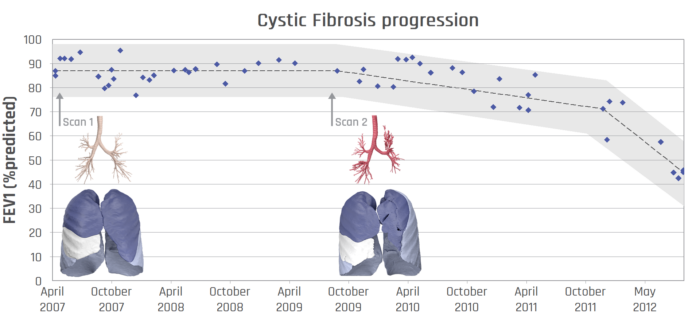
Traditional lung function measurements, such as FEV1 or FVC, only provide meaningful information about the condition of the entire lung and do not indicate where the exhaled air is coming from.
Regional information is often crucial to understand the pathophysiology of the individual patient and to give guidance for optimal treatment.
Functional Respiratory Imaging (FRI) is a clinically meaningful and non-invasive measurement of the patient-specific respiratory system. A set of distinct biomarkers analyzes exposure, structure and function of the lungs and airways in any respiratory disease.
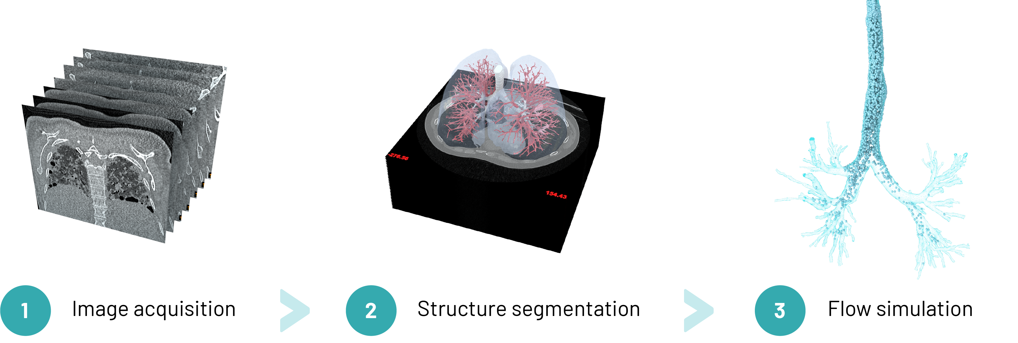
The FRI process consists of three phases:
PHASE 1: medical imaging
The process starts with the acquisition of low dose, high-resolution computed tomography (HRCT) scans of the patient.
PHASE 2: Image Processing
Measurements are performed on the segmented 3-dimensional geometries from these scans.
PHASE 3: Flow Simulation
Computational Fluid Dynamics (CFD) is used to quantify airflow and exposure to inhaled particles.
This results in a number of important biomarkers which are most relevant in therapy development and patient care. The usage of FRI biomarkers is scalable and easy to implement.
Airway resistance
Air trapping
Ventilation mapping
Ventilation / Perfusion reserve
Lung and lobar volume
Emphysema
Internal airflow distribution
Blood vessel volume
Nodule volume

Airway (wall) volume

Aerosol deposition
Why regional information matters
Traditional lung function measurements, such as FEV1 or FVC, only provide meaningful information about the condition of the entire lung and do not indicate where the exhaled air is coming from.
Regional information is often crucial to understand the pathophysiology of the individual patient and to give guidance for optimal treatment.
Sensitive biomarkers which measure lung disease stage and the biological response to treatment are needed to facilitate clinical development decisions, accelerate drug development and improve patient care.
Therefore, FRI is an essential addition to the toolkit of research and clinical practice in respiratory diseases.
Fluidda’s goal is
to help people suffering
from respiratory diseases
get the treatment they need
85% accuracy
in predicting Bronchiolitis Obliterans Syndrome (BOS)
< 6 weeks
is the average time in which clients receive their results after having delivered all input data
Up to 16 times less
patient enrolment needed to establish significant result with FRI
More accurate data
to strengthen your drug administration procedure
Our responsibility
At Fluidda, we strive to continuously increase our knowledge, to continuously improve our imaging parameters, and to continuously innovate. In doing so, we aim to provide the respiratory field (drug developers, medical device developers, researchers, clinicians) with the most meaningful, quantitative and efficient imaging solutions. Fluidda is very conscious of the potential harm that exposure to high radiation doses can cause when patients undergo CT scans. Therefore, we take every precaution to minimize radiation exposure for each subject through tailored and qualitative scanning protocols, smart trial designs, strict scanner quality checks, and trainings at the scanning sites. We strongly support CT scanner innovations to limit doses even further.
We are always available if you wish to receive more information regarding radiation dosage and FRI.
Healthcare company?
Healthcare company?
Discover how Fluidda can speed up your respiratory drug or medical device development and clinical trial.
Healthcare provider?
Healthcare provider?
Fluidda assists radiologists and pneumologists to optimize the respiratory patient’s treatment.
News & events
News & events
Want to follow our work? Read all about our latest news and events. Check out our social media.

