
Functional Respiratory Imaging can predict the course of COVID-19
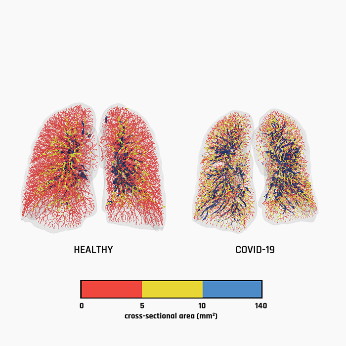
Functional Respiratory Imaging (FRI) quantifies and visualizes the distribution of blood vessels in the lungs in great detail. This simple model, which combines the results of the imaging with the patient’s age, can accurately predict the risk of dying and the necessity for ventilation.
Clinical research conducted has revealed that inflammation of the endothelium and microangiopathy occur commonly in the blood vessels of the lungs of people who died because of aSARS-CoV-2 infection. This observation led to the hypothesis that visible changes in the perfusion of the lungs on a CT scan could be a biomarker for a poor prognosis with COVID-19. Important changes in perfusion were visible in the blood vessels with a cross-sectional surface area of 1.25 to 5 mm2.
A group of American researchers led by Prof. Mike Morris and Prof. Marilyn Goldberg, tested this hypothesis during a retrospective study using high-resolution CT images.
This study revealed that BV5 was significantly reduced in COVID-19 subjects compared to healthy individuals, and it was found to be related to the severity of COVID-19 infection. FRI has the potential to standardize and quantify the analysis of CT images of the lung at a level that not every radiologist can achieve. For COVID-19, FRI is proving to be useful as part of a simple clinical decision model.
To read the full article, click HERE.
Categorised in: Articles, Miscellaneous / January 18, 2022 2:51 pm /
Tags: COVID19, FRI, Magazine