
Airway volume measurement with FRI identifies IPF progression
Idiopathic pulmonary fibrosis (IPF) is a progressive disease with unclear etiology.
IPF patients decline with heterogeneous trajectories. The ability to determine an individual’s disease course is limited to pulmonary function measurement (pulmonary function testing (PFT)) and visual assessment of computed tomography (CT) scans, both of which have limitations. PFT is affected by patient technique and operator experience. CT scans rely on a radiologist’s expertise to correctly interpret disease stability/progression and are subject to inter-observer variability.
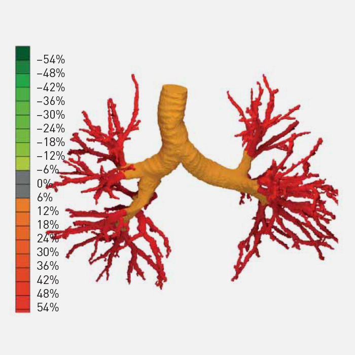
A recent publication shows that airway volume measurements performed with Functional Respiratory Imaging (FRI) can discriminate between patients with stable and progressive IPF at the time the CT scans were taken. This technique can be very useful, for instance, in clinical trials where new antifibrotic drugs are being investigated.
Categorised in: Research / February 25, 2020 10:48 am /
Tags: Clinical, IPF, Trials