
New FRI Article: Cluckers et al: Interstitial lung disease in systemic sclerosis
FRI is able to differentiate moderate to severe versus limited disease and to detect disease progression in systemic sclerosis.
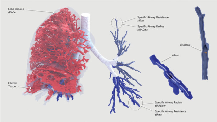
Systemic sclerosis–associated ILD accounts for up to 20% of mortality in these patients and has a highly variable prognosis. Functional respiratory imaging (FRI), a quantitative computed tomography imaging technique which allows mapping of regional information, can provide a detailed view of lung structures. It thereby shows potential to better characterize this disease.
In a recently published study in the Journal of Scleroderma and Related Disorders, an observational cohort of 35 patients with systemic sclerosis was retrospectively studied by comparing serial pulmonary function tests and in- and expiratory high-resolution computed tomography over 1.5year interval. After classification into moderate to severe lung disease and limited lung disease, post hoc analysis was performed using mixed-effects models and estimated marginal means in terms of functional respiratory imaging parameters.
It was concluded that FRI—in terms of lower lobe volumes, siRaw, and siRADaw—can differentiate between moderate to severe and limited diseases, predict disease progression, and is able to detect short-term changes in SSc-ILD, independent of disease severity. These novel findings should be prospectively validated in earlier disease and other SSc-ILD cohorts, in conjunction with other measures of disease progression.
Categorised in: Miscellaneous / January 28, 2021 1:38 pm /
Tags: FRI, Sclerosis